A 2016 comparison study found CBCT scans changed the implant treatment plan in up to 45% of cases, versus a panoramic X-ray alone (PubMed 26227397, Clinical Oral Implants Research). That’s a striking number for a scan patients are often told is “just a precaution.”
Most clinics mention a CBCT scan without explaining what it’s actually looking for. This guide covers what the scan shows, how it changes real treatment decisions, what it costs in Dubai, and when it’s genuinely necessary rather than routine upselling.
Key Takeaways
- CBCT changed the implant treatment plan in roughly 28-45% of cases compared to panoramic X-ray alone (PubMed 26227397, 2016).
- It measures bone density in Hounsfield Units, classifying implant sites from dense (D1) to soft (D4), which determines implant size and whether grafting is needed.
- The scan itself takes 10-40 seconds, at roughly a fifth the radiation of a medical CT scan.
What Is a CBCT Scan?
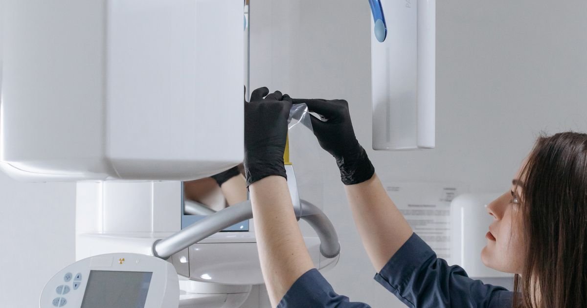
A CBCT (cone-beam CT) scan builds a 3D image of your jaw in a single 10-40 second rotation, capturing bone, nerves, and sinus structures a flat 2D X-ray simply can’t show. The machine rotates a cone-shaped X-ray beam around your head once, and software reconstructs the data into a 3D model your dentist can rotate and measure on screen.
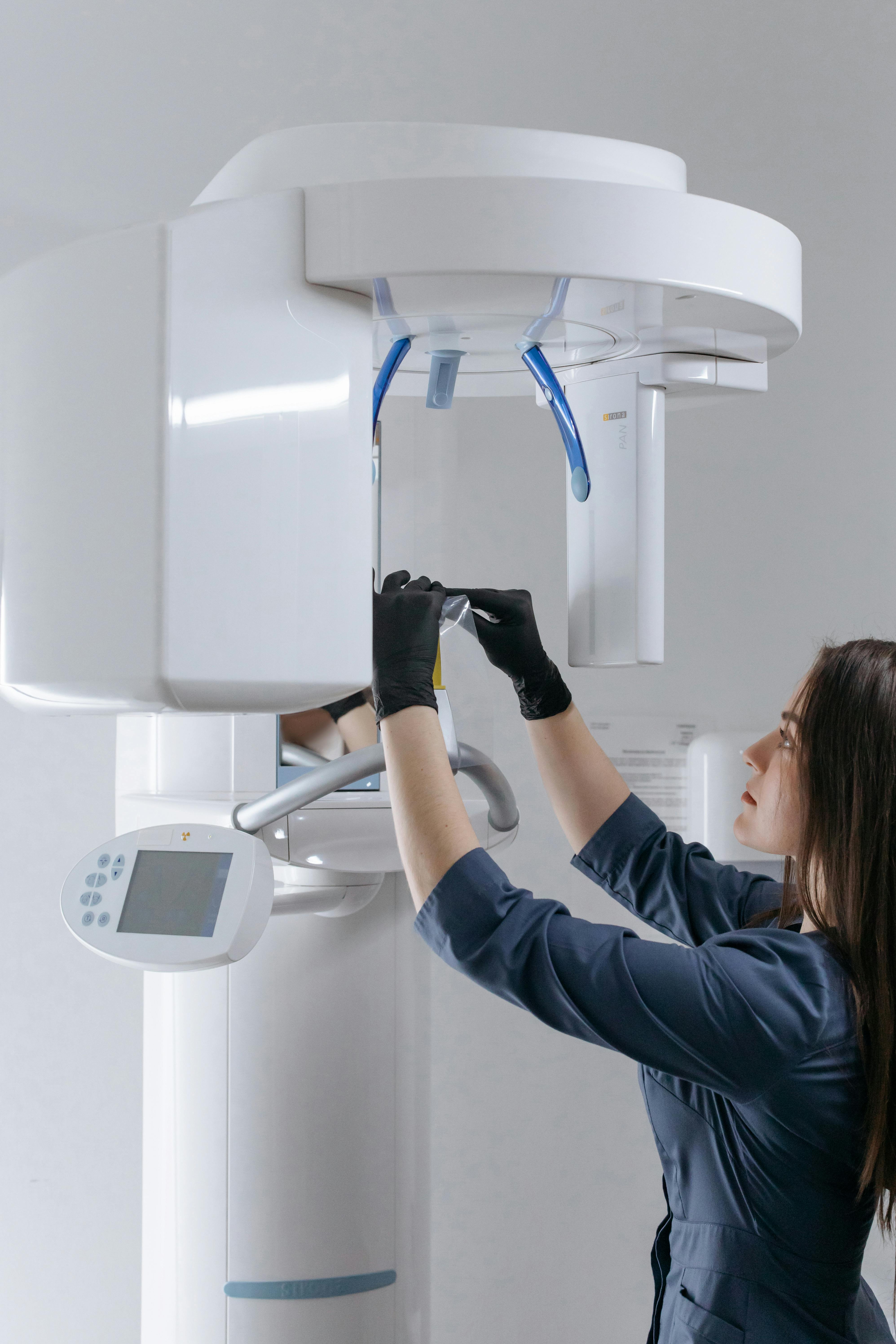
A panoramic X-ray, by contrast, flattens everything into one 2D image, which is fine for spotting cavities but leaves depth and width as guesswork. A medical CT scan captures similar 3D detail to CBCT, but at a much higher radiation dose, more machine than dentistry actually needs. The full appointment, including positioning, usually runs 5-10 minutes.
For the complete digital implant workflow, see Digital Dental Implants in Dubai.
What Does a CBCT Scan Actually Show for Implant Planning?
A CBCT scan measures bone density in Hounsfield Units (HU) and classifies implant sites from D1 (dense, above 1,250 HU) to D4 (soft, 150-350 HU), a classification that directly determines implant length, width, and whether grafting is needed (PMC10485442, 2023).
D1 bone is common in the front of the lower jaw and handles implant placement predictably. D4 bone shows up most often in the back of the upper jaw, where it’s softer and heals more slowly, which is exactly why posterior maxilla implants tend to need more planning, sometimes including a bone graft before the implant goes in at all.
Bone density is only one input, though. Two other structures decide whether an implant site is straightforward or complicated: the sinus and the nerve.
For how this bone and anatomy data becomes a physical surgical guide, see In-House 3D Printing: Why It Speeds Up Implant Treatment.
How Does CBCT Map the Sinus and Nerve Before Surgery?
A 450-patient CBCT study found root protrusion into the maxillary sinus in 41.3% of cases, most often at the first molar, with sinus-floor-to-crest distance ranging from just 1.2mm to 4.2mm depending on tooth position (PMC12859313, 2025). At 1.2mm of available bone, a sinus lift stops being optional.
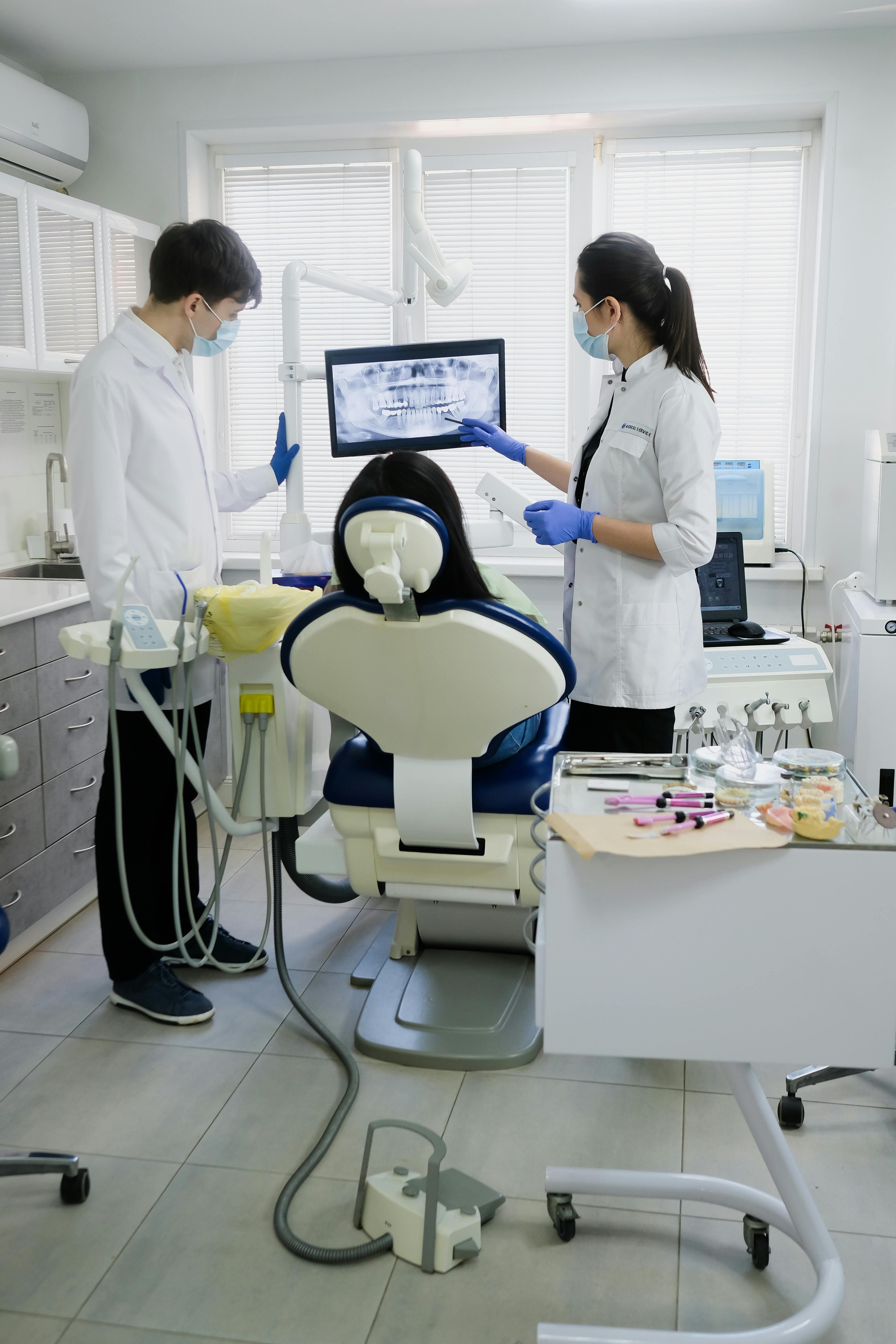
The same scan maps the inferior alveolar nerve canal in the lower jaw. Placing an implant too close to that nerve risks numbness or altered sensation, a complication that’s entirely preventable once the canal’s position is known in 3D rather than estimated from a flat image. The same study found sinus septa present in 62.7% of cases, another detail invisible on a panoramic X-ray.
[PERSONAL EXPERIENCE] In one case at our clinic, a patient referred for a routine upper premolar implant turned out to have a sinus floor sitting noticeably lower than the panoramic image had suggested. The CBCT scan caught it before surgery, not during it, which is the entire point of scanning in 3D rather than 2D.
Does CBCT Change the Treatment Plan Compared to a Regular X-ray?
A comparison study found CBCT and panoramic X-ray plans agreed in only 55-72% of cases, meaning CBCT altered the surgical plan in roughly 28-45% of cases, mostly toward more conservative approaches or added grafting (PubMed 26227397, 2016). Panoramic X-rays consistently overestimated available bone height, particularly before decisions about vertical bone grafting.
Worth flagging honestly: this comparison traces back to a single 2016 study. No one appears to have re-run it more recently, which says as much about the industry’s evidence base as it does about CBCT itself. It’s still the best available data, so we’re citing it, but “best available” isn’t the same as “current-year.”
Is a CBCT Scan Safe? Dose, Field of View, and Special Cases
CBCT delivers roughly a fifth the radiation of a medical CT scan, and 99.7% of dental CBCT scans use a small or medium field of view (FOV), the setting that keeps dose as low as reasonably achievable (EADMFR dose-optimization survey). Isn’t it worth asking your clinic which FOV they plan to use before your scan?
Field of view is simply how much of your jaw the scan captures. A limited FOV, under 8cm, covers just the implant site. A medium FOV, 8 to 15cm, captures more of the arch. A large FOV goes further still, but for planning a single implant, it’s rarely necessary. Smaller FOV means less tissue exposed and a lower dose, without losing the detail that matters for your specific case.
As for pregnancy: there’s no absolute contraindication, but it’s handled as a risk-benefit decision with lead shielding, and most clinics avoid non-urgent imaging as a precaution regardless (IAEA radiation protection guidance).
For the full radiation dose comparison, see the CBCT safety section of Digital Dental Implants in Dubai.
What to Expect During a CBCT Appointment in Dubai
A CBCT scan requires no special preparation beyond removing metal jewelry, and typically costs AED 500-800 for a single jaw in Dubai, though pricing varies by clinic and isn’t usually covered by basic UAE dental insurance. Treat that figure as a general range rather than a fixed quote, costs shift by clinic and by whether one or both jaws are scanned.

[UNIQUE INSIGHT] Most Dubai clinic pages either quote a price with no context or avoid pricing entirely. Neither is that useful. What actually matters is asking directly: does this quote include both jaws, and is it billed separately from the consultation? Basic UAE dental insurance plans typically classify implant-related imaging as elective, so confirming coverage with your insurer before booking saves a surprise later.
Frequently Asked Questions
How much does a CBCT scan cost in Dubai?
Typically AED 500-800 for a single jaw and AED 700-1,000+ for both jaws, though pricing varies by clinic. Treat this as a general range and confirm directly with your provider before booking.
Is a CBCT scan covered by dental insurance in the UAE?
Usually not under basic plans, since imaging tied to implant treatment is often classified as elective. Check directly with your insurer, coverage varies significantly between providers.
Can a CBCT scan detect infection or cysts before implant placement?
Yes. CBCT’s 3D detail can reveal cysts, infections, and bone abnormalities that a 2D X-ray may miss entirely, which is part of why it changes treatment plans in a meaningful share of cases (PubMed 26227397, 2016).
How long does a CBCT scan take?
The scan itself captures in 10-40 seconds, with the full appointment, including positioning and instructions, typically running 5-10 minutes.
Is a CBCT scan safe during pregnancy?
There’s no absolute contraindication, but it’s handled as a risk-benefit decision with lead shielding, and most clinics avoid non-urgent imaging during pregnancy as a precaution (IAEA guidance).
What’s the difference between a CBCT scan and a regular dental X-ray?
A regular dental X-ray is a flat 2D image at a very low dose. CBCT builds a 3D model showing bone density, nerve position, and sinus anatomy, detail a 2D X-ray simply cannot capture.
[INTERNAL-LINK: what the scan determines about your candidacy → Am I a Good Candidate for Dental Implants? full eligibility guide]
Conclusion
A CBCT scan isn’t a formality before implant surgery. It’s the reason a plan changes in roughly a third of cases, catching sinus proximity, nerve position, and bone density issues a panoramic X-ray would miss. Understanding what your scan actually measures, in Hounsfield units, in millimeters of sinus clearance, in field-of-view size, turns an opaque step into an informed decision.
If you’re planning implant treatment, ask specifically what field of view your clinic uses and whether the scan is done in-house. Those two answers tell you more than any marketing page will.
For how this scan data feeds into surgical guide accuracy, see 3D Guided Implant Surgery: How Surgical Guides Improve Accuracy.
[INTERNAL-LINK: why imaging affects your treatment plan and price → Dental Implant Cost in Dubai pillar page]
Sources
- PMC, Clinical Assessment of Bone Quality at Implant Site using CBCT and Hounsfield Unit, retrieved 2026-07-07, 2023
- PMC, 3D Imaging of Maxillary Sinus Anatomical Variations Using CBCT, retrieved 2026-07-07, 2025
- PubMed, Implant Treatment Planning: Panoramic Radiographs vs. CBCT, Clinical Oral Implants Research, retrieved 2026-07-07, 2016
- IAEA, Radiation Protection of Pregnant Women in Dental Radiology, retrieved 2026-07-07